
How to Immediately Feel Better
The establishment calls it “Hormone Replacement”, but the word “replacement” contains a concession. It implies that something has been lost permanently and must be substituted from outside. Testosterone replacement therapy. Thyroid replacement. Hormone replacement therapy. The language of replacement accepts decline as a fait accompli and offers a pharmaceutical prosthetic.
Restoration is a different proposition. It assumes the system is intact, the pathways are present, the substrate is available — and that what has declined can be maintained or returned to functional levels through intervention that works with the body’s own mechanisms rather than substituting for them. The distinction is not semantic; it determines the entire therapeutic strategy and most of its outcomes.
This chapter is about restoration.
A hormone is a signaling molecule produced in one location and transported through the bloodstream to act on distant target tissues. What distinguishes hormones from other biochemical actors is their role as master regulators — each one binding to receptors in cells throughout the body and altering gene expression across dozens or hundreds of downstream targets simultaneously. Cortisol doesn’t do one thing. It modulates immune function, glucose metabolism, bone density, memory consolidation, skin integrity, and cardiovascular tone in parallel, through a single molecular signal. Vitamin D — misclassified as a vitamin, functioning as a steroid hormone — regulates over 200 genes in cells ranging from immune tissue to neurons to cardiac muscle. Testosterone doesn’t build muscle; it activates the genetic programs that build muscle, maintain libido, regulate red blood cell production, protect bone density, and sustain cognitive function, all at once. This is why the endocrine cascade’s decline with age produces effects that appear across every organ system simultaneously — not because aging is attacking everything independently, but because the master regulators governing everything are running down together. The aging clock hypothesis follows directly from this architecture: if hormones regulate gene expression system-wide, and if hormonal levels decline with age in predictable patterns, then hormonal decline is not merely a symptom of aging. It is one of its primary drivers.
The endocrine system does not fail all at once. It winds down through a cascade of interrelated declines, each one reducing the substrate or signal for the next, across decades — in ways that medicine has generally treated as separate conditions requiring separate prescriptions rather than as a single integrated system requiring integrated support. The goal of the protocol described here is to maintain the entire cascade, simultaneously, through the same portfolio approach that governs every other dimension of this book.
The Cascade Begins With Cholesterol
Everything in this chapter starts with cholesterol. Not the dietary villain of thirty years of flawed nutritional policy but the substrate for every steroid hormone the human body produces.
The pathway is linear and elegant: dietary and endogenously produced cholesterol is converted in mitochondria to pregnenolone, the grandmother molecule of all steroid hormones. Pregnenolone then branches into DHEA, which branches into testosterone, estrogen, and progesterone. It also branches into cortisol and aldosterone through a separate arm. Every hormone discussed in this chapter is downstream of that single conversion step from cholesterol.
This is why the cholesterol-lowering imperative of mainstream cardiology collides so destructively with the hormonal landscape of aging men. The substrate for testosterone is the same molecule being pharmacologically suppressed. The substrate for DHEA, for pregnenolone itself, for every steroid the body uses to maintain its endocrine function, is the molecule medicine has spent four decades telling people to reduce at any cost.
As the Workout chapter will document, the cholesterol that was previously being converted into steroid hormones begins to accumulate in the bloodstream when testosterone production declines in the late forties. The correct reading of rising cholesterol in a late-middle-aged man is not necessarily “cardiovascular risk requiring a statin.” It may be “reduced downstream demand for the substrate as hormonal production winds down.” The statin that then suppresses the cholesterol also suppresses what remains of the steroid synthesis capacity. The iatrogenic acceleration runs in both directions.
Vitamin D: The Hormone Nobody Calls a Hormone
Vitamin D is not just a vitamin. It is a steroid hormone precursor synthesized from cholesterol, which is the same cholesterol that begins the pregnenolone cascade. The designation “vitamin” is a historical accident of nomenclature that has caused incalculable harm by implying that adequate dietary intake or modest supplementation is sufficient, when the evidence shows consistently that it is not.
The Life Extension Foundation conducted an analysis of 13,000 blood tests of dedicated supplement users in 2009. These were not ordinary health consumers. They were people taking supplements daily, aware of vitamin D’s importance, actively trying to maintain their health. The results showed that 85.7% had insufficient 25-hydroxyvitamin D blood levels below the minimum protective threshold of 50 ng/mL. Three years later, after awareness had dramatically increased and supplement doses had risen substantially, a follow-up analysis of 39,925 blood test results found that 63.8% were still below optimal levels.
The magnitude of this deficit in a highly motivated, supplement-taking population tells you something important about the general population not taking supplements at all.
The optimal range in the blood is 50-80 ng/mL of 25-hydroxyvitamin D. The consequences of falling below it are not subtle. Insufficient vitamin D is associated with virtually every age-related disorder: cancer, cardiovascular disease, chronic inflammation, neurodegeneration, autoimmune disease, respiratory infection, osteoporosis, and metabolic dysfunction. The mechanisms have been characterized: vitamin D receptors are present in virtually every cell type, and calcitriol, the active hormone form, regulates over 200 genes involved in cell proliferation, immune surveillance, inflammation modulation, and calcium metabolism.
At this point, many might wonder if those levels are “unnaturally high”. They aren’t. Anyone out in the sun on a summer day naturally produces far higher amounts of Vitamin D. The problem for our modern world is that most people never spend enough time in the sun! One hour in the sun generates about 30,000 IU of Vitamin D. Darker skinned individuals of course produce less Vitamin D with sun exposure because melanin blocks the critical UVB light.
A gold standard double-blind, controlled study published in the American Journal of Clinical Nutrition found that women given supplemental vitamin D and calcium had a greater than 50% reduction in cancer risk over a four-year trial compared to placebo. That is the same magnitude of effect as the selenium NPC trial. Two separate vitamins, two separate mechanisms, two separate randomized controlled trials stopped or producing results so dramatic they could not be ignored — and the mainstream clinical response to both has been to minimize, contextualize, and wait for pharmaceutical confirmation that will never arrive because neither compound can be patented.
The practical details matter and are rarely communicated. Aging persons require approximately 5,000 IU per day to achieve optimal blood levels — roughly ten times the typical American intake. The dose must be taken with the heaviest meal of the day: a Cleveland Clinic Foundation study demonstrated that taking vitamin D with the largest meal increased blood levels by over 50% compared to taking it on an empty stomach. Vitamin D is fat-soluble, and fat in the meal is required for absorption. A health-conscious person reducing their fat intake while taking vitamin D may be inadvertently creating the deficiency they are trying to prevent.
The thyroid connection is direct. Vitamin D receptors regulate genes involved in thyroid hormone synthesis, and vitamin D deficiency is associated with increased risk of Hashimoto’s thyroiditis — the autoimmune condition that is the most common cause of hypothyroidism in the developed world. Managing one supports the other.
The cholesterol connection closes the circle: vitamin D is synthesized from 7-dehydrocholesterol in skin exposed to UVB radiation, a pathway that becomes dramatically less efficient with age. Young skin produces abundant vitamin D from sun exposure. Aging skin does not. Studies of older adults who spend substantial time outdoors in Hawaii found surprisingly low vitamin D blood levels. The sun is there. The capacity to use it is diminished. The supplement is not optional — it is what aging biology requires to compensate for what aging skin can no longer produce.
Thyroid: What the Blood Tests Revealed
Some vulnerabilities announce themselves early. My mother took thyroid medication for her entire adult life. That family history meant the question was not whether I might develop thyroid dysfunction but when, and whether I would catch it before it had done significant damage.
It appeared in routine blood testing which I obtain at vast discount through the Life Extension Foundation. The kind of systematic monitoring that most physicians perform at annual checkups might not contain the needed blood tests to catch an early decline. The TSH was elevated beyond the upper reference range, and the free T3 was at the low end of normal. Subclinical hypothyroidism by conventional definition, because the T4 appeared adequate. Not subclinical in any functional sense.
The thyroid system has a similar problem to the cholesterol-testosterone relationship: the standard clinical measurement is inadequate. TSH, Thyroid Stimulating Hormone, is what the pituitary produces to tell the thyroid to make more hormone. An elevated TSH means the pituitary is shouting at the thyroid. A normal TSH means the shouting has achieved the desired result. What TSH does not tell you is whether the T4 being produced is efficiently converting to T3 — the active hormone form that actually enters cells and regulates metabolism, body temperature, heart rate, cognitive function, and energy production. T4 is the storage form. T3 does the work.
The conversion of T4 to T3 requires deiodinase enzymes. These enzymes are selenium-dependent — which means that selenium, already in the protocol for its cancer-preventive properties documented in Chapter 3, is also essential for thyroid hormone activation. A person with adequate T4 and inadequate selenium may have effectively reduced T3 despite normal-appearing thyroid function tests. The iodine required to build T4 in the first place — four iodine atoms per T4 molecule, three per T3 — must also be adequate, and iodine deficiency remains surprisingly common despite iodized salt programs.
My approach follows the same principle as every other chapter in this book: no pharmaceuticals if a natural intervention exists and the data supports it. The natural thyroid support protocol includes iodine and selenium as the essential cofactors for hormone synthesis and conversion, zinc as an additional deiodinase cofactor, and tyrosine — the amino acid from which thyroid hormones are built. Two tyrosine molecules linked together, with iodine atoms attached: that is what T4 is. Providing the building blocks supports the system’s own production.
Ashwagandha, already present in the cognitive stack through Cognitex Elite, has documented thyroid-supporting properties. It has been shown to stimulate thyroid hormone synthesis and increase both T3 and T4 levels in clinical studies, operating through HPA axis normalization rather than direct thyroid stimulation. A supplement taken for cortisol management is simultaneously supporting thyroid function. That is the portfolio approach demonstrating its efficiency: multiple mechanisms from single interventions, chosen because they address independent targets that turn out to be more connected than they appear.
The monitoring imperative is the chapter’s methodological contribution: blood testing caught what symptom-watching would have missed for years. Low-grade hypothyroidism is a slow thief. Fatigue dismissed as aging. Cognitive slowing attributed to stress. Cold sensitivity written off as a personal quirk. By the time the symptoms become unmistakable, the deficit has been running for a decade. A full thyroid panel — TSH, free T3, free T4, and ideally reverse T3 — run annually provides the data that symptom-watching cannot.
A Critical Warning Before the Protocol
The thyroid supplement market contains a hazard that most consumers never suspect: products labeled as natural herbal thyroid support that secretly contain pharmaceutical levothyroxine or desiccated thyroid tissue. This is not speculation. It is documented in the medical literature, including a peer-reviewed case report published in the American Journal of Case Reports describing a 70-year-old man who developed thyroid storm — a life-threatening emergency requiring intubation, cardioversion for atrial fibrillation, and 17 days of intensive care — after taking a Chinese herbal product that laboratory analysis confirmed was contaminated with levothyroxine. The paper cites additional prior cases of thyroid tissue found in Chinese herbal weight loss products specifically. The FDA has taken enforcement action on multiple “natural” thyroid support supplements found to contain undisclosed thyroid hormones.
The implication is straightforward: most off-brand thyroid support products are not what they claim to be. The “natural” product producing measurable thyroid effects may be producing those effects because it contains pharmaceutical-grade thyroid hormone never disclosed on the label. For someone who chose natural intervention specifically to avoid pharmaceutical dependency, this is the most fundamental violation possible — and it carries real risk. A person with subclinical hypothyroidism taking an undisclosed levothyroxine dose can swing into hyperthyroidism with consequences ranging from arrhythmia and bone loss to the thyroid storm documented in the case report.
The solution is not to avoid thyroid support. It is to understand exactly what each ingredient is doing and why.
The Validated Protocol
After extensive trial and elimination — discarding products that produced effects inconsistent with their labeled ingredients — my current thyroid restoration protocol rests on four components with characterized mechanisms and documented safety profiles.
Sea Iodine and Potassium Iodide (KI) provide dual-form iodine supplementation. Thyroid hormone synthesis is impossible without iodine: T4 contains four iodine atoms, T3 contains three. Sea iodine derived from algae provides molecular iodine (I₂), while potassium iodide provides iodide (I⁻). Different tissues preferentially utilize different forms: the thyroid uses iodide for hormone synthesis, while other glandular tissues use molecular iodine. The combination ensures adequate substrate across all iodine-utilizing tissues. This is the foundational intervention; provide what the thyroid needs to produce hormone rather than stimulating a gland that lacks raw material.
Schisandra (Schizandra chinensis, Wu Wei Zi) addresses the thyroid system from two independent angles. Its active schisandrin compounds are potently hepatoprotective — protecting the liver enzymes that perform approximately 60% of the body’s T4 to T3 conversion via type 1 deiodinase. A liver under oxidative stress loses deiodinase efficiency, producing functional T3 deficiency regardless of how much T4 the thyroid generates. This is one mechanism by which chronic stress, environmental toxins, and inflammatory states produce hypothyroid symptoms despite normal-appearing gland function. Schisandra also normalizes the HPA axis — elevated cortisol directly impairs thyroid function at multiple levels — and protects thyroid tissue from oxidative damage directly. One herb, three independent thyroid-relevant mechanisms, none involving exogenous hormone.
Forskolin (from Coleus forskohlii) is the most pharmacologically specific intervention in the protocol. Forskolin directly activates adenylyl cyclase, the enzyme producing cyclic AMP — the same secondary messenger that mediates TSH’s stimulating effects on thyroid follicular cells. Forskolin amplifies that intracellular signal, reinforcing the TSH message at the cellular level. Clinical studies confirm that forskolin supplementation increases both T3 and T4 serum levels. It also activates cAMP-mediated lipolysis in adipose tissue which directly addresses the weight gain tendency that is the most functionally significant consequence of subclinical hypothyroidism. The same compound supports thyroid hormone production and promotes fat mobilization through an independent pathway simultaneously.
Ashwagandha (Withania somnifera) is already running in the protocol through Cognitex Elite for cortisol management and cognitive support, but its thyroid effects deserve explicit recognition here rather than a footnote. A randomized double-blind placebo-controlled trial published in the Journal of Alternative and Complementary Medicine found that ashwagandha root extract supplementation significantly increased serum T3 and T4 levels in subjects with subclinical hypothyroidism over eight weeks. The mechanism is not direct thyroid stimulation but rather HPA axis normalization: chronic cortisol elevation directly suppresses thyroid function at multiple points — inhibiting TRH release from the hypothalamus, reducing TSH sensitivity at the thyroid, and impairing T4 to T3 conversion peripherally. By normalizing cortisol through withanolide-mediated adrenal modulation, ashwagandha removes a systematic brake on thyroid function that stress physiology imposes.
This is the portfolio approach’s characteristic efficiency made visible. Ashwagandha appears in the cognitive chapter for its cortisol-neuroprotection effects, in the testosterone chapter for its HPA axis support, and now in the thyroid protocol for its cortisol-mediated thyroid support — three chapters, one supplement, three independent mechanisms pointing in the same direction. It was always doing all three simultaneously. The protocol captured the cognitive benefit first; the thyroid benefit was running in parallel before it was formally recognized.
Ashwagandha called a “panacea” is not hyperbole. It reflects the reality that the HPA axis connects to virtually every endocrine system, and a compound that normalizes the HPA axis therefore has downstream effects across virtually every hormone discussed in this chapter. Ashwagandha is not doing many things. It is doing one thing, normalizing the central stress response system, with positive effects through the entire endocrine cascade.
The Down-Regulation Problem — Why Replacement Defeats Itself
There is a deeper reason to avoid exogenous thyroid hormone beyond the contamination risk and the vegetarian objection to desiccated animal glands. It is the same reason testosterone patches are the wrong intervention for low testosterone, and the same reason this chapter is titled Restoration rather than Replacement.
The hypothalamic-pituitary-thyroid axis is a closed-loop control system. When thyroid hormone circulates at adequate levels, the hypothalamus reduces its TRH signal and the pituitary reduces its TSH output — the gland is told it can rest. Under normal circumstances this is elegant regulation. When the source of that circulating thyroid hormone is external rather than endogenous, the regulatory signal is identical but the consequence is destructive: the gland receives progressively less stimulation, reduces its own output, and over time atrophies from disuse.
The PMC contamination case documents this outcome explicitly. When the endocrinologist evaluated the patient — a known hypothyroid man who had been on levothyroxine for years before the herbal contamination episode — they declined to add propylthiouracil because the patient likely had an atrophic thyroid from long-term exogenous hormone use. The organ had essentially retired. Years of pharmaceutical replacement had not maintained thyroid function. They had replaced it with a dependency while the gland progressively lost the capacity to function independently.
This is the strategic error that replacement therapy makes at every level of the endocrine system. Testosterone replacement suppresses the hypothalamic-pituitary-gonadal axis, reduces LH and FSH, and causes testicular atrophy. Exogenous growth hormone suppresses endogenous GH secretion. Every introduced hormone tells the body’s own production apparatus that its services are no longer required.
The protocol described in this chapter does the opposite at every step. Iodine provides substrate that requires the gland to actively synthesize hormone. Forskolin amplifies the TSH signal inside thyroid cells, making the gland more responsive to its own regulatory inputs rather than bypassing them. Schisandra supports the conversion pathway so that whatever the gland produces is efficiently activated downstream. The axis stays intact. The gland stays exercised. The feedback loop keeps running.
Support the system’s own production where a suppressible feedback loop makes substitution self-defeating. Substitute directly where structural decline has removed the production capacity that the feedback loop once regulated. That is the principle that distinguishes restoration from replacement — stated in the thyroid system with particular clarity, applicable across every hormone discussed in this chapter.
The primary residual symptom of the hereditary thyroid tendency — in the presence of the full protocol — is a disposition toward weight gain rather than its realization. Untreated subclinical hypothyroidism produces measurable weight accumulation over years through reduced basal metabolic rate, impaired fat oxidation, reduced thermogenesis, and water retention. None of these are present at clinical levels despite the underlying genetic vulnerability.
The protocol is not eliminating the thyroid tendency. It is neutralizing its metabolic consequences sufficiently to maintain the hormonal output and conversion efficiency that the genetics would otherwise erode. The blood tests confirm the intervention is working. That is the only confirmation that matters.
Potential Alternatives
Maca represents a class of adaptogens that modulates the function of hormonal systems without significantly changing hormone levels. Studies in ovariectomized rats showed maca balancing effects on estrogen, progesterone, cortisol, ACTH, and thyroid hormones simultaneously, with additional antidepressant-like effects — suggesting modulation of the hypothalamic-pituitary axis rather than direct hormone production. Clinical studies confirm improved sexual desire, erectile function, and menopausal symptom relief without measurable hormone level changes. Its bioactive compounds — macamides, macaenes, and glucosinolates — contribute neuroprotective, anti-inflammatory, and antioxidant effects alongside the hormonal modulation. (Ulloa del Carpio, 2024). Hopefully more studies will be done in the future to identify the mechanism.
When Down-Regulation Does Not Apply
The down-regulation argument requires an important qualification. It applies most forcefully to hormones regulated by tight pituitary negative feedback loops — the HPG axis governing testosterone, the HPT axis governing thyroid hormones, and the HPA axis governing cortisol. For these systems, exogenous hormone directly suppresses the pituitary signals that maintain endogenous production, and the producing gland atrophies from disuse over time.
Not all hormonal decline follows this architecture. For several hormones central to the longevity protocol, the decline is primarily structural rather than regulatory — and the feedback mechanisms that would produce down-regulation either do not exist or are too loose to create meaningful dependency.
Melatonin is the clearest case. The pineal gland physically calcifies with age — a structural deterioration documented on CT scans, not a functional suppression by feedback signaling. Melatonin production is regulated by the suprachiasmatic nucleus circadian clock and darkness exposure, not by a pituitary feedback loop that exogenous melatonin could suppress. The gland is producing less because it has calcified, not because it detects adequate circulating levels and reduces output accordingly. Supplementing melatonin replaces what a structurally compromised gland cannot produce. The natural alternatives are limited precisely because the problem is structural: tart cherry juice provides small dietary melatonin quantities, and strict light discipline — complete darkness at night, bright morning light — supports whatever circadian signaling capacity remains. Neither meaningfully replaces the supplement. Melatonin is the legitimate exception to the restoration-not-replacement philosophy.
Pregnenolone synthesis from cholesterol in mitochondria is regulated by StAR protein activity and mitochondrial function rather than by pituitary feedback. Supplementing pregnenolone does not suppress endogenous synthesis the way testosterone supplementation suppresses the HPG axis. The age-related decline is primarily substrate and mitochondrial-capacity limited — the same mitochondrial atrophy that the pre-workout stack addresses — rather than feedback-loop limited. Some monitoring of downstream conversion is warranted: supplemented pregnenolone under chronic stress conditions may be preferentially shunted toward cortisol production, shifting the hormonal balance in ways that periodic testing would reveal.
DHEA is produced primarily by the adrenal zona reticularis under ACTH stimulation, but the feedback regulation is substantially looser than the testosterone or thyroid axes. The age-related decline in DHEA is driven primarily by adrenal zona reticularis atrophy — again structural — rather than altered feedback signaling. Some modest suppression of endogenous DHEA production occurs with supplementation, but it does not produce the dependency and glandular atrophy seen with direct sex hormone replacement. DHEA also serves as a precursor that the body converts according to its own downstream needs, making it a substrate intervention as much as a replacement.
The practical principle is this: support endogenous production through upstream intervention wherever a suppressible pituitary feedback loop governs the system. Supplement directly where the decline is structural and the feedback architecture is absent or loose enough to make dependency unlikely. Test periodically to confirm the intervention is producing the intended result without creating downstream imbalances that testing alone would reveal.
The protocol already embodies this distinction. Thyroid and testosterone receive upstream substrate and cofactor support — iodine, selenium, the aromatase inhibitors, the SHBG competitors. Melatonin, pregnenolone, and DHEA receive direct supplementation. The philosophy is the same. The application differs because the biology demands it.
The Declining Roster
Beyond thyroid and the testosterone pathway described above, the endocrine system loses ground across multiple independent fronts simultaneously. Addressing any one of them without addressing the others misses the cascade structure of the problem.
This Is Aging
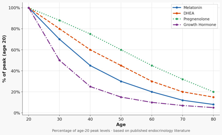
Pregnenolone is the first loss — the grandmother molecule whose decline reduces substrate availability for everything downstream. Levels begin declining in the late twenties and continue throughout life. Supplementing pregnenolone directly provides the starting material for the entire steroid cascade, including neurosteroid synthesis in the brain, where pregnenolone has independent cognitive effects.
DHEA peaks in the early twenties and declines approximately 2% per year thereafter. By age seventy, levels are 10-20% of peak values. DHEA is the most abundant circulating steroid hormone in humans and the immediate precursor to both testosterone and estrogen. It also has direct effects on immune function, insulin sensitivity, cardiovascular protection, and bone density independent of its role as a sex hormone precursor. The 7-Keto DHEA metabolite in the pre-workout stack addresses a different downstream position — the non-hormonal metabolite that supports metabolism without converting to sex hormones — while direct DHEA supplementation maintains the precursor pool.
Melatonin is the most visibly functional decline because its consequences are immediately experiential: sleep architecture deteriorates, the deep slow-wave sleep required for glymphatic waste clearance from the brain diminishes, and the downstream cognitive and recovery effects accumulate nightly. The pineal gland calcifies progressively with age, producing melatonin at a fraction of young-adult levels by the seventh decade. Supplementing melatonin is not creating dependency — it is compensating for a physically diminished gland. The dose question is important: the pharmaceutical convention of 10mg doses is almost certainly excessive. Low doses in the 0.5-3mg range more closely replicate physiological nocturnal peaks and avoid the receptor desensitization that high doses may produce. A time-release tablet more closely matches the natural release throughout the night.
Growth Hormone / IGF-1 decline constitutes what endocrinologists call the somatopause — the gradual suppression of the pulsatile nocturnal growth hormone secretion that peaks in deep sleep and drives the tissue repair, muscle protein synthesis, and metabolic regulation that distinguish a young body from an old one. By sixty, pulsatile GH output is roughly 25% of young adult levels. Natural stimulation of GH secretion is achievable through specific amino acids — arginine and ornithine taken before sleep, glycine in similar fashion — and through the vigorous resistance training that is the most reliable natural GH secretagogue available. What the pre-workout chapter documents for other reasons turns out to extend this considerably: Alpha-GPC stimulates GH secretion by inhibiting hypothalamic somatostatin through cholinergic pathways, with one study recording 44-fold GH increases when combined with exercise; CDP-choline operates through a parallel somatostatin-inhibiting mechanism and has been shown to double the GH response to GHRH in elderly subjects aged 69-84; and acetyl-L-carnitine amplifies downstream IGF-1 signaling that translates GH into tissue effects. The pre-workout stack is hitting GH secretion from five independent angles without having been assembled with that explicitly in mind — a convergence the chapter on that stack documents in full.
Progesterone declines in both sexes, a fact that receives almost no attention in male health discussions. Progesterone is neuroprotective, promotes GABA receptor sensitivity producing calming effects, modulates the immune system, and counterbalances the estrogenic effects that aromatization of testosterone produces. Its decline in aging men is one of the underappreciated contributors to the anxiety, sleep disruption, and inflammatory background that mainstream medicine addresses with pharmaceuticals rather than with the hormone whose absence is driving the symptom. Pregnenalone supplementation can enable more progesterone but not necessarily. Blood tests for it are relatively expensive, but a progesterone cream is cheap and widely available. If you notice improvements in sleep or handling stress, keep with it.
The Aging Clock Hypothesis
We do not know what drives the aging clock. The honest answer to that question is that nobody does, and the confident assertions of researchers who claim to have found the single mechanism — telomere attrition, epigenetic methylation drift, mitochondrial dysfunction, senescent cell accumulation, stem cell exhaustion — are each identifying real phenomena while almost certainly missing the full picture.
What the hormonal framework offers that the others don’t is a systems-level explanation for why so many of these phenomena appear simultaneously. If hormones are master regulators — each one altering gene expression across dozens or hundreds of downstream targets at once — then a coordinated decline in the hormonal cascade would produce coordinated downstream failures across every system those hormones regulate. Telomeres shorten faster in tissues with declining growth hormone and IGF-1 signaling. Epigenetic methylation patterns drift in cells deprived of adequate progesterone and estrogen. Mitochondria dysfunction accelerates as thyroid hormone declines and the metabolic rate they depend on for maintenance signals drops. Senescent cells accumulate faster as DHEA and its downstream immune modulators fall away. The other theories are not wrong. They may be describing the downstream consequences of the same upstream failure — the master regulators losing their grip — rather than independent causes competing for primacy.
The hormonal theory of aging has an underappreciated champion in Vladimir Dilman, the Soviet physician-scientist who proposed in the 1950s that the hypothalamus progressively loses sensitivity to hormonal feedback signals as it ages — raising the threshold required to trigger hormonal responses and causing the entire cascade to run at progressively lower set points. On Dilman’s model, the body is not running out of the capacity to produce hormones. The control system is losing its calibration. The thermostat is drifting, not the furnace.
The TRIIM trial provides the most striking evidence that hormonal restoration affects the aging clock itself rather than merely its symptoms. Healthy men treated with growth hormone, DHEA, and metformin for one year showed a 2.5-year reversal on Horvath’s epigenetic DNA methylation clock which is a validated biological age measure that is currently the best available proxy for the true aging rate. The hormonal intervention didn’t just slow aging; it moved the clock backwards.
If the epigenetic clock is the aging clock, and if hormonal decline is one of its primary drivers, then maintaining the full hormone cascade is not symptomatic treatment. It is intervention at the mechanism — the same ambition that drives the senolytic protocol, the mitochondrial support, and the neurite regeneration program, addressed through the endocrine system rather than the cellular system.
The chapter on combining interventions will address what happens when you run all these interventions simultaneously. The present observation is narrower: the hormonal restoration protocol is not a cosmetic effort to feel younger. It is maintenance of the signaling architecture that governs how quickly every other system ages.
The Monitoring Imperative
None of this works without data. The portfolio approach to hormonal restoration requires periodic blood testing to confirm that the interventions are producing the intended results, to catch what symptom-watching misses, and to adjust doses as the years accumulate and the systems change.
The minimum useful panel for a man pursuing hormonal restoration includes: 25-hydroxyvitamin D, TSH with free T3 and free T4, total and free testosterone, DHEA-S, IGF-1 as a proxy for growth hormone output, estradiol, and a comprehensive metabolic panel that captures thyroid, liver, and kidney function simultaneously. Homocysteine and a complete lipid fractionation — not just total cholesterol but LDL particle size, HDL, triglycerides — close the picture.
I found out with a blood test that even the modest 50 mg of DHEA I was taking had built up to greatly excessive levels, so I switched to the DHEA metabolite. Whatever the rest of my protocol is doing, it apparently has already kept my DHEA levels from declining.
This is not medicine as most people experience it. It is research conducted on the most relevant subject available: yourself. The ABA methodology described elsewhere in this book applies here: establish a baseline, intervene, measure again, compare. Adjust based on data rather than on symptoms alone.
The blood tests are the instrument. The protocol is the experiment. The decades are the sample size.
None of this requires a physician’s order, a referral, or an insurance authorization. Direct-to-consumer laboratory testing services — Life Extension Foundation’s annual blood test sale being the most comprehensive and cost-effective — allow anyone to order the full panel described above, walk into a LabCorp collection center, and receive results within days. The same tests that cost $2,000 or more when ordered through a physician’s office are available for a fraction of that price ordered directly. The LEF cost even includes a physician’s consultation if desired. The monitoring imperative does not require the medical gatekeeping system. It requires the decision to look.
My thyroid deficiency would still be progressing unchecked, at a pace that symptom-watching alone would not have revealed for years, if routine testing had not made the invisible visible. The natural intervention addressed it, and the monitoring proved it worked.
That is the methodology of this entire book applied to one hormone in one system. Multiply it across the full cascade and you have the practice of restoration rather than the acceptance of replacement.
Leave a Reply